Biomedical Engineering Reference
In-Depth Information
In this case, we segment an artery from an 155
×
170
×
165 image volume
obtained from the Visible Man Project. The T-Surfaces parameters are:
c
=0
.
75,
k
=1
.
12,
γ
=0
.
3, grid resolution 4
×
4
×
4, and freezing point set to 10. The
result of steps (1)-(6) is depicted in Figure 11a.
Among the proposals to address this problem (relax the threshold, mathemati-
cal morphology [33], etc), we have tested anisotropic diffusion [32]. The properties
of this method (see Appendix A) enable smoothing within regions in preference
to smoothing across boundaries. Figure 11b shows the correct result obtained
when pre-processing the image with anisotropic diffusion and then applying steps
(1)-(6).
6.4. CT Images
In this section we test our approach for a 150
×
175
×
180 computed to-
mography image. The structure of interest is the right kidney. Figure 12a shows
the result of steps (1)-(4) when using
T
∈
(205
,
255) in Eq. (7) as well as the
following parameters:
γ
=0
.
01,
k
=1
.
222, and
c
=0
.
750. The total number of
evolutions was 8. The grid resolution is 3
×
3
×
3.
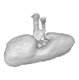
Like in previous examples, the genus of the extracted surface is not the correct
one and some small disconnected components appear (Figure 12a). After 10
iterations of the T-Surfaces algorithm, the geometry extracted becomes that one of
the target. Figures 12b and 12c show that the defects (holes) observed in Figure 12a
have disappeared and that the final topology is the correct one.
(a)
(b)
(c)
Figure 12.
(a) Result for steps (1)-(4) for CT volume images. (b) View of final solution
showing that topological defects were corrected. (c) Another view of the final solution.