Biomedical Engineering Reference
In-Depth Information
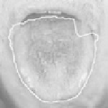
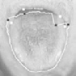
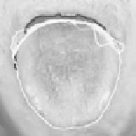
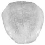
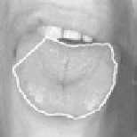
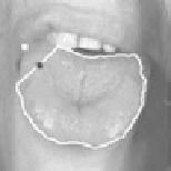
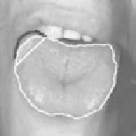
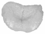
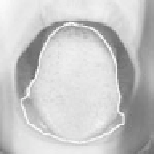
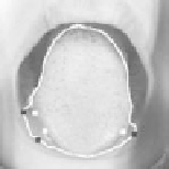
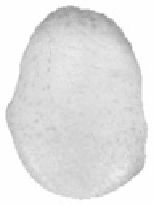
Figure 8.
Examples of interactive segmentation on three tongue images. The four columns
from left to right are: original segmentation results from the color GVF snakes, point pairs
chosen for the snake pit, convergence of the snakes, and final extracted tongue bodies.
See
attached CD for color version.
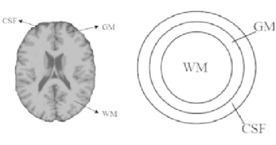
Figure 9.
Left: transversal slice of MR brain volume constituting WM, GM, and CSF, as
labeled. Right: simplified model with ribbon-like structure. While real cerebral cortex has
varying thickness, it is approximated with constant thickness in the simplified model. The
motivation for this simplification will be discussed in more depth below.