Biomedical Engineering Reference
In-Depth Information
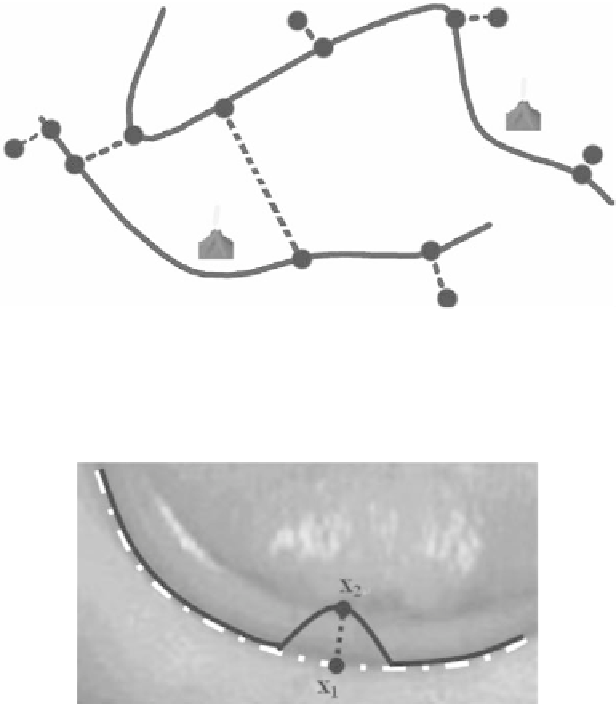
Figure 6.
Geometric interpretation of Snake Pit. The two dark curves are different snakes,
and the tow springs (dashed line) are connected between them to create a coupling effect.
The other springs attach points on the snakes to fixed positions in the image. In addition,
two volcanos are set to bend a nearby snake.
See attached CD for color version.
Figure 7.
A pair of points for the snake pit.
See attached CD for color version.
5.
CEREBRAL CORTEX MR IMAGE SEGMENTATION
5.1. Introduction
The cerebral cortex is a thin but convoluted layer of neurons known as Gray
Matter (GM) that lies between the White Matter (WM) and the Cerebral Spinal
Fluid (CSF) (Figure 9). This layer is only 1-3 millimeters thick and has a total
area of about one quarter of a square meter. It is by far the largest portion of the
nervous system in the human body. Studies show that many neurological disorders
like Alzheimer's disease and schizophrenia are directly related to the pathological
state of the cerebral cortex. Therefore, extraction of cerebral cortex from the brain
for analysis and measurement is necessary and important.