Biomedical Engineering Reference
In-Depth Information
(a)
(b)
(c)
(d)
(e)
(f)
(g)
(i)
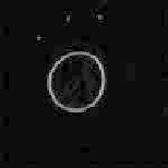
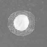
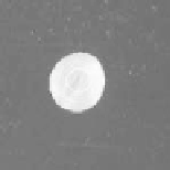
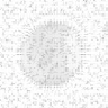
Figure 3.
An example of segmenting a seed image using a color GVF snake: (a) original
color seed image; (b,c) filtering results from Gaussian kernel and anisotropic diffusion,
respectively; (d) edge map using a Di Zenzo color gradient operator; (e,f) convergence of
two color GVF snakes with different initial curves; (g,h) closeups of external force fields in
a color GVF snake and a classical snake, respectively.
See attached CD for color version.
4.
TONGUE SEGMENTATION BASED ONA COLOR GVF SNAKE
It seems unreasonable to directly apply the proposed color GVF snake model
to tongue image segmentation. There are a lot of pathological details on the
surface of the tongue (e.g., tongue fur) with ambiguous colors, cracks, petechia,
and highlight areas, which have a strong influence on the convergence of the snake.
Although we have adopted the anisotropic diffusion technique to filter a tongue
image, sometimes the snake curve still falls into the local minimum. To overcome
these problems, a great deal of prior knowledge has been considered to provide an
automatic curve initialization method for the color GVF snake.
Generally, in the captured tongue images (see Figures 4a-c), the following
classes of colors are contained in the challenging areas: yellow (face skins), white
(teeth), black (shadow produced by lips), etc. On the other hand, the color of lips
is often red, and the substrate color of the tongue is red as well. According to this
observation, we can alleviate the effects of most of the challenging areas first. The
following transform of the original tongue image is used for this purpose:
I
(
x, y
)=
|
R
(
x, y
)+
B
(
x, y
)
−
2
G
(
x, y
)
|
,
(25)
where
R
(
x, y
),
G
(
x, y
), and
B
(
x, y
) are the three components of the color image.
This transform may effectively enhance the contrast between the tongue body and