Biomedical Engineering Reference
In-Depth Information
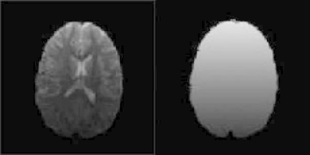
Figure 1.
Intensity inhomogeneities in brain MR imaging.
segmentations allow for uncertainty in the location of object boundaries, where
voxels may be classified into multiple classes with a varying degree of member-
ship. The membership thus gives an indication of where partial-volume effects
have occurred in the image.
Nonuniformity of regional intensities is a major difficulty that is specific to the
segmentation of MR images [6,7]. It results in slow intensity variations of the same
tissue over the image domain. The sources for this include poor radio-frequency
(RF) coil uniformity, static field inhomogeneity, RF penetration, gradient-driven
eddy currents, and overall patient anatomy and position [6]. This variability of
tissue intensity values with respect to image location can significantly affect vi-
sual evaluation and degrade the performance of methods which assume that the
intensity value of a tissue class is constant over the image. Recently, a number
of image processing solutions [8-11] have been proposed as well as extended to
simultaneously correct for the bias field and segment MR brain images in [12].
Figure 1 gives an example of intensity inhomogeneities in brain MRI.
1.1.2. Priors
In many applications, computer processing and analysis of medical images is
limited due to low contrast of anatomical structures, variability of tissue features,
fuzziness of boundaries between different tissues, and distributional complexities
of tiny structures such as veins and nerves. In an effort to overcome these diffi-
culties, various techniques have been developed to incorporate prior information
into the segmentation process. When segmenting or locating an object, prior in-
formation — such as the general shape, size, location, and orientation of an object
— can be quite helpful in improving the efficiency and accuracy of segmentation.
Prior-based image segmentation that incorporates prior information of a certain