Biomedical Engineering Reference
In-Depth Information
(a)
(b)
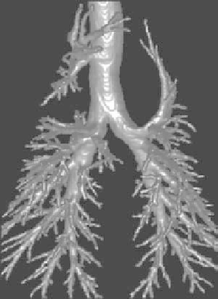
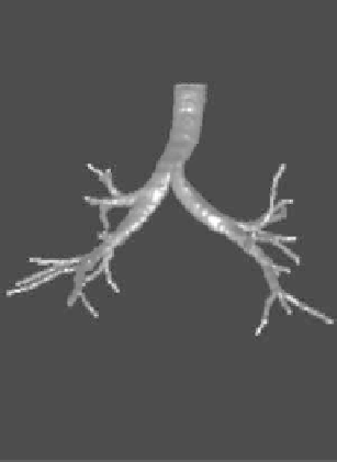
Figure 6.
Results of pulmonary airway tree segmentation using 3D Fast MarchingMethods
from 3D CT image data: (a) pulmonary airway tree of a sheep lung from 3D CT data; (b)
pulmonary airway tree of a human lung from low-dose 3D CT data.
as a tool to prevent leakage. An example is shown in Figure 7a,b. The size of this
dataset is 319
382. The processing time using the Fast Marching Meth-
ods is less than 20 seconds. The high proceeding speed due to the Fast Marching
Method is very valuable in clinical usage. A modified version of this method was
applied to measurement of coronary vasoreactivity in sheep using 64-slice multi-
detector computed tomography and 3d segmentation [44]. Some results are shown
in Figure 7c,d.
One similarity of the above examples is that the boundary of the object to be
segmented is easy to find. That is why the simple speed factor is defined based on
intensity or as a function of intensity. However, inmost cases, especially inmedical
image processing, medical image segmentation often faces difficult challenges,
including poor image contrast, noise, and missing or diffuse boundaries.
×
400
×
3.3. Other Applications
To determine the minimum cost path after the endpoint is reached, a backprop-
agation from the endpoint to the starting point is carried out [45]. In the isotropic
marching case, the fastest traveling is always along the direction perpendicular to
the wave front, i.e., the iso-curve of the arrival time. Therefore, the minimum cost
path can be found by a gradient descent in the arrival time function. A simple