Chemistry Reference
In-Depth Information
(a)
(b)
5000
4000
3000
2000
1000
0
50
100
150
200
STRUCTURE NUMBER
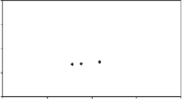
Figure 5.15
(A) COST versus rank for the NOE matching run on Bcl-x
L
/
7
poses using the
protein coordinates from 1LXL. The poses of the NOE matching run are ordered based on
COST and plotted from lowest COST to highest COST. (B) Superposition of target pose and the
minimum cost pose (dark gray) from (A). The side-chains of F97, Y101 and R139 from 1YSG
(light gray) and 1LXL (dark gray) are also displayed.
In the second test case, we used the Bcl-x
L
coordinates from the apo X-ray structure
(1MAZ). The RMSD of backbone atoms involved in secondary structure of 1MAZ is
1.39 Å from 1YSG and 1.54 Å from 1LXL, the NMR apo structure. Trial binding poses
of
7
were generated with
Poser
using a binding site box set to ligand binding site plus an
additional1Åinallcoordinate axes and a 5° rotational sampling. Over 734 000 poses
were evaluated, with 53 797 poses being retained by
Poser
. To reduce the calculation
time while we optimize the code for dealing with larger number of poses, 10 000 poses
were randomly selected from the
Poser
ensemble for evaluation with NOE matching. NOE
matching was applied using our 57 experimental intermolecular NOEs and with the pre-
dicted protein resonance assignments set to the corresponding BMRB average values. The
COST values from NOE matching ranged from 1429 to 4429 (Figure 5.16A). Cluster-
ing of the poses based on RMSD and a similarity of 0.8 Å resulted in 522 clusters, of
which 122 were singletons. As indicated above, comparison with the poses in each of
the clusters to the pose for
7
found in the Bcl-x
L
/
7
complex is complicated by the struc-
tural differences between the apo and bound protein structures. Superimposing the protein
backbone atoms involved in secondary structure and then calculating the RMSD for res-
ultant positions of
7
in each of the structures, the pose with the lowest COST has an
RMSD of 3.19 Å with the target pose. For the ensemble of
Poser
trial poses, RMSDs
to the target pose ranged from 1.40 to 8.33 Å. As mentioned above, several residues
in the binding pocket of apo Bcl-x
L
to adopt different conformations upon
7
binding
(Figure 5.16B).
In the third test case, we used the Bcl-x
L
coordinates from the NMR structure of the
complex BAK-Bcl-x
L
(1BXL). The RMSD of backbone atoms involved in secondary
structure of 1BXL is 1.53 Å from 1YSG. Trial binding poses of
7
were generated with
Poser
using a binding site box set to ligand binding site plus an additional 1 Å in all
coordinate axes and a 5° rotational sampling. Over 795 000 poses were evaluated, with
127 289 poses being retained by
Poser
. To reduce the calculation time while we optimized