Biomedical Engineering Reference
In-Depth Information
18
140
16
14
120
12
100
10
80
8
6
60
4
40
2
20
0
0
2
4
6
8
10
12
14
16
18
20
0
2
4
6
8
10
12
14
16
18
20
(a) (b)
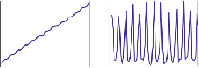
Fig. 10.22.
Pf-RBC displacement (a) and velocity (b) along the wall for the schizont stage (from
[51])
time (s)
time (s)
small contact area with the wall. During these steps Pf-RBCs undergo strong mem-
brane deformations as illustrated in Fig. 10.21. A similar behaviour was found in
experiments [81] of Pf-RBCs which showed flipping (rolling) along a wall coated
with purified ICAM-1. In agreement with the simulations, RBCs in experiments also
showed strong membrane deformations characterized by local membrane buckling.
Fig. 10.22 presents the corresponding displacement along the
x
coordinate (a)
and instantaneous RBC velocity (b). An infected RBC rolls in a relatively stable
motion which resembles a staircase. The segments of smaller displacements corre-
spond to the stage of a flat RBC adhesion followed by its slow peeling off the wall
(see Fig. 10.21), while the fragments of larger displacements represent the stage
of RBC fast flipping described above. The RBC velocity is in agreement with its
displacement showing high peaks or fast cell motion during the time segments with
larger displacements. The average cell velocity is approximately 5
m/s. Fig. 10.23
shows RBC displacement along the
z
cross-flow coordinate (a) and instantaneous
contact area (b). The displacement across the wall shows a jerky motion of an in-
fected RBC within several microns. This is due to the discrete number of bonds and
their random rupture or dissociation. Thus, if there is a non-uniform distribution of
.
8
μ
10.5
50
45
10
40
9.5
35
9
30
8.5
25
8
20
7.5
15
7
10
0
2
4
6
8
10
12
14
16
18
20
0
2
4
6
8
10
12
14
16
18
20
(a) (b)
Fig. 10.23.
RBC displacement across the wall (a) and the cell contact area (b) for the schizont stage
(from [51])
time (s)
time (s)