Biomedical Engineering Reference
In-Depth Information
where
1
λ
2
t
λ
=
C
0
:
H
0
,
H
0
=
κ
I
+(
1
−
3
κ
)
a
0
⊗
a
0
,
2
a
π
0
ρ
(
Θ
)
1
4
sin
3
κ
=
Θ
d
Θ
.
(6.40)
ρ
(
Θ
)
Material parameters
λ
a
,
a
0
and
can be obtained from fibre images and used
,
η
to calculate
H
0
and
can be obtained from the nonlinear
stretch-stress data. It is straightforward to consider an integral fibre representation
as in (6.26) and (6.29) rather than the GST model [50], or to use planar splay rather
than conical splay.
Samples of left common carotid arteries were obtained from New Zealand white
rabbits and tested in the UA-MPM device (Fig. 6.4) using a previously developed
protocol [49, 50]. Briefly, a sample of artery was removed from its source, opened
longitudinally, manually cut into circumferential strips with a “dogbone” shape, and
then placed in the UA-MPM system for extension tests. Prior to testing, the thickness
(H) and width (W) of the unloaded tissue sample were measured five times with
calipers and averaged to obtain cross sectional area.
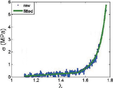
After preconditioning, force and stretch values were recorded under quasi-static
loading conditions, Fig. 6.5. Force data recorded from the load cell was converted
to Cauchy stress assuming an isochoric deformation. Stretch
κ
. Constants
η
and
γ
iso
was measured di-
rectly from displacement in the uniaxial device. For a given deformation of the ar-
terial strip, we then obtained averaged Cauchy stress as a function of sample stretch
Fig. 6.5. The collagen fibres were imaged at six different stretch values during this
process Fig. 6.6.
λ
Fig. 6.5.
Raw and fitted data from uniaxial extension of Sample 01 of rabbit carotid artery. Corre-
sponding MPM-UA images are shown in Fig. 6.6