Biomedical Engineering Reference
In-Depth Information
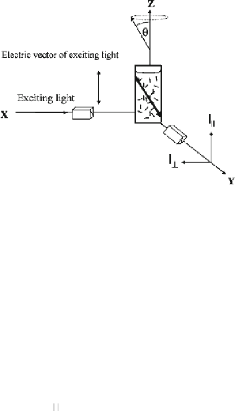
Fig. 9.2
System for
measuring anisotropy.
The
z
-axis is the vertical
laboratory axis. The sample
is placed at the origin, and
readings are taken along the
y
-axis. Reprinted with
permission from Jameson and
Ross (
2010
) . Copyright 2010
American Chemical Society
can be manipulated experimentally by rotating the polarizer. Figure
9.2
shows how
these principles can be applied experimentally to measure the fluorescence anisot-
ropy of a sample. In this case, a polarizer is inserted in a beam of light directed along
the
x
-axis, allowing the isolation of an electric vector oriented parallel to the
z
-axis.
When this polarized light strikes the sample at the center of the coordinate system,
the fluorescently tagged molecules in solution that have an appropriately oriented
absorption transition dipole moment will become excited and fluoresce. The inten-
sity of fluorescence emission is measured 90° to the axis of excitation, along the
y
-axis, in the planes parallel and perpendicular to the
z
-axis. The relationship between
these intensities gives a measure of the mobility of the fluorescently tagged probe.
In this system, anisotropy (
A
) is defined as the ratio of the linearly polarized
component of emitted light over the total intensity or
I
−
I
⊥
A
=
(9.1)
I
+
2
I
⊥
= 0, giving
A
= 1), it means that the probe is immobile since excitation and emission are both in
the same plane. Conversely, if the probe rotates at infinite speed,
I
ïï
will be equal to
I
Theoretically, when all emission is parallel to the
z
-axis (
I
ïï
= 1 and
I
⊥
, yielding
A
= 0. Thus,
A
is expected to fall between 1 and 0 for most commonly
used fluorophores under solution conditions, indicating complete polarization and
depolarization, respectively. Under some circumstances, it is possible to observe
A
< 0, although this requires that the population of emission dipoles be heavily
biased in the plane perpendicular to excitation, limiting where
I
ïï
= 0 and
I
⊥
= 1, giv-
ing
A
= −0.5. However, the high and low extremes of anisotropy are normally only
encountered in highly structured samples such as crystals and not normally observed
in solution-based biochemical assays (Jameson and Ross
2010
) .
⊥

Search WWH ::

Custom Search