Biology Reference
In-Depth Information
B
A
D
C
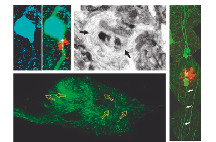
Figure 5.2. Partial blockage of axonal transport in amyloid or tau pathology. (A) Axons (green)
swell next to amyloid plaques (red), and rounded mitochondria (blue) accumulate in
these swellings. (B) Microtubule whorls (arrows) are evident in some swellings.
(C) Similar microtubule diversions (green EB3 staining) form in Aplysia when tau is
overexpressed (see also supplementary movie in Shemesh et al., 2008). (D) Despite the
extensive swelling, axons in the mouse amyloid model shown in A and B remain
continuous and morphologically normal at more distal sites (white arrows) for several
months, indicating continued flow of at least some transport. A, B, and D reproduced
from Adalbert
et al.
(2009) with permission from Oxford Journals. C reproduced from
Shemesh
et al.
(2008) with permission from John Wiley & Sons, Inc.
that in young rats and may decline by as much as 71% (Frolkis
, 1997;
Minoshima and Cross, 2008). Similar events occur in older primates (Kimura
et al.
et al.
, 2007). Until noninvasive methods can be applied in man, we can
only guess how much more transport slows over the course of 80 years or
more. As axonal transport impairment is clearly established as a cause of some
neurodegenerative conditions (Martin
, 2002), this age-
related decline could predispose to a range of age-related disorders or even cause
some.
et al.
, 2002; Reid
et al.
Thus, young axons appear to transport far more material than they need
for survival, because otherwise, an age-related decline would cause massive axon
death. This could explain the survival of swollen axons in young amyloid mouse
models (Adalbert
, 2005; Fig. 5.2). If the defect were
imposed on a lower basal level of transport, the consequences might be far worse
et al.
, 2009; Spires
et al.