Biology Reference
In-Depth Information
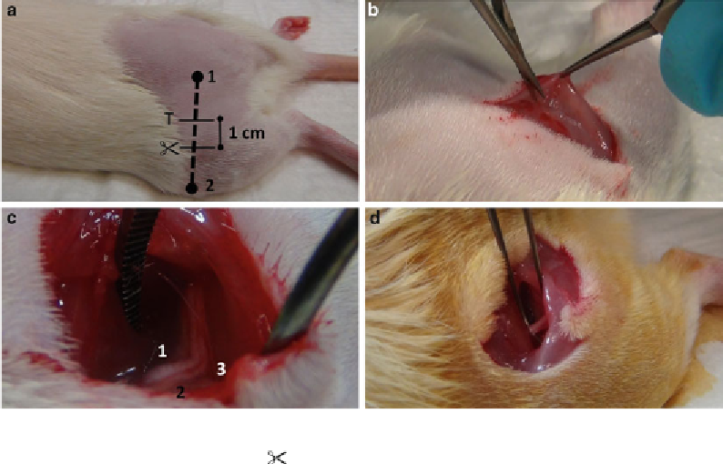
Fig. 1
Surgery for induction of the spared nerve injury. (
a
) Superficial landmarks for orientation: (1) Crest of
ilium, (2) Patella, (T) Site of trifurcation, ( ): Location of fi rst incision. (
b
) Making the incision in between the
two heads of the biceps femoris muscle with a micro scissor to enter the site of the location where the trifurca-
tion is being situated. (
c
) Sciating nerve and trifurcation: (1) Common peroneal nerve, (2) Tibial nerve, (3) Sural
nerve. (
d
) Ligation and transection of the common peroneal nerve. Lifting the nerve produces a bridge that
allows safe transection of the nerve
11. Insert the Metzenbaum scissors horizontally and closed into
the small incision between the skin and the muscle layer and
detach the skin from the underlying tissue by opening the scis-
sors and carefully withdrawing it. Repeat this procedure until
the skin is sufficiently detached.
12. Make an incision to proximal with a total length of 3-4 cm
following the femoral bone.
13. Retract the skin to expose the underlying muscles.
14. Locate the margins of the two heads of the biceps femoris
muscle, which is characterized by a white line of adjoining
fascia.
15. Carefully lift the medial part of the muscle with the Bonn
micro forceps to create a small indentation (Fig.
1b
).
16. Carefully cut the fascia with the Vannas spring scissors to
detach the muscles. This allows the exposure of the space
where the nerves and vessels are situated.
17. Expose the sciatic nerve and its trifurcation carefully by blunt
preparation with the standard pattern curved forceps. Insert
the forceps in a closed manner and allow it to open in order to
make space (see Note 2). Be careful not to touch or stretch the
Search WWH ::

Custom Search