Biology Reference
In-Depth Information
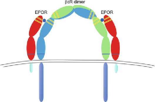
Fig. 1
The innate repair receptor. Available evidence indicates that the receptor
mediating tissue protection and repair is assembled from subunits consisting of
EPOR and
β
CR (for review, see ref.
19
)
fractions prepared from tissues exhibiting tissue protection (e.g.,
brain, kidney, liver) and were subsequently passed over the column
(
16
). After extensive washing, the retained proteins were removed
from the column, run on a gel and probed with anti-EPOR or
anti-
CR
and EPOR subunits were among the proteins retained on the
CEPO column. Confirmation that the beta
β
CR antibodies. The results clearly showed that both
β
CR was indeed a
component of the tissue protective receptor was confirmed by
studying mice in which the
β
CR was knocked-out. Although these
mice are phenotypically normal while young, as predicted their
tissues do not respond to either EPO or non-erythropoietic tissue
protective molecules following injury (
16
). Based on the biology
of the related GM-CSF receptor, the proposed structure of the
tissue protective receptor is illustrated in Fig.
1
(reviewed in ref.
19
).
The signaling pathways activated by EPO in tissue protection
include the same JAK2-signal transducer and activator of tran-
scription (STAT) utilized in erythropoiesis. However, multiple
alternative pathways have also been identified, including the pro-
tein kinase B-Akt system, as well as mitogen-activated kinases
(reviewed in ref.
19
).
β
5
Exogenous EPO for Tissue Protection?
Although many acute experimental models show impressive pro-
tective effects following administration of exogenous EPO, there
are major potential roadblocks for clinical utility. Clearly, the use
of systemically administered EPO for tissue protection requires a
Search WWH ::

Custom Search