Agriculture Reference
In-Depth Information
AP
VP
W
E1
E1
E1
E2
10 cm
E2
E2
E3
E3
10 min
50 mV
E3
_
E4
+
E4
E4
_
+
W
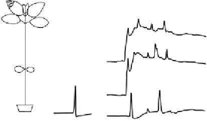
Fig. 19.1.
Action potentials (
APs
)andvariationpotentials(
VPs
) recorded in the stem of
Helianthus annuus
by extracellular electrodes,
E1
-
E4
. The AP was elicited by electrical
stimulation (
), and the VP by wounding (
W
).
Ve r t i c a l a r r o w s
indicate the moment of
stimulation.
Arrowheads
point to the direction of propagation (After Stankovic et al. 1998)
±
phase is followed by slower repolarizing and after-hyperpolarizing phases.
APs can be evoked by relatively weak, nondamaging stimuli. The same
place can be stimulated many times without visible damage. APs spread on
the basis of local electrical circuits.
19.1.2
Ion Mechanism of Action Potentials
The ion mechanism of APs was intensely studied in giant
Characean
cells.
The huge dimensions of internodal cells enable application of many exper-
imental techniques, including voltage-clamp, patch-clamp with a patch-
pipetteattachedtotheplasmamembranethrougha“window”cutinacell
wall, and calcium imaging with aequorin or fura microinjected into a cell.
Thecellscanbesurgicallymodifiedbycuttingoffthenodesandsubsequent
exchange of the vacuolar sap and even tonoplast decomposition (Tazawa
and Shimmen 1987).
In resting cells Ca
2+
and Cl
−
are kept far from the electrochemical equi-
librium. These two ion species are the best candidates as depolarizing ions.
Indeed, according to the generally accepted model, an AP is initiated by
calcium influx into the cytosol followed by Cl
−
efflux (Williamson and
Ashley 1982; Lunevsky et al. 1983). Chloride ions leave the cell down their
electrochemical potential gradient through Ca
2+
-activated anion channels.
Repolarization occurs after opening of voltage-gated potassium channels of


Search WWH ::

Custom Search