Biology Reference
In-Depth Information
Haversian canal
D
Lacuna
O
M
(a)
(b)
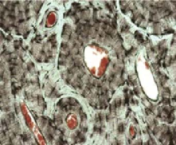
Figure 4.6
Cellular material in bones and teeth. (a) cross-section through a femur: the Haversian
canals are surrounded by concentric layers of bone (lamellae); bone cells (osteocytes) occupy
lancunae (Image provided by Prof Tim Arnett, Department of Cell & Developmental Biology,
University College London, UK). (b) cross-section through a human tooth showing the dentine
(D), odontoblast layer (O) and middle part of the dental pulp (M) (Image provided by Dr Marko
Vavpotic, Institute of Forensic Medicine, University of Ljubljana, Slovenia)
(a)
(b)
(c)
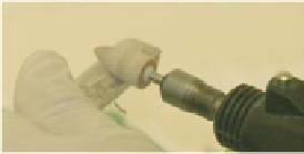
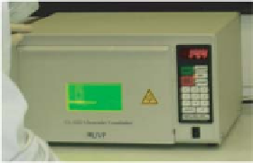
Figure 4.7
Bone and tooth material can be vigorously cleaned using: (a) abrasion to remove
the outer surface and (b) washing in detergent and bleach to remove contaminating materials.
(c) Exposure to strong UV light introduces thymine dimers into any contaminating exogenous
DNA - preventing amplification during PCR
detergents to remove any soft tissue [33], followed by physical abrasion, soaking in
sodium hypochlorite (bleach) [34] (Figure 4.7), trypsin enzyme [35] and exposure
to strong ultraviolet light.
After cleaning, the bone/tooth material is normally broken down into a powder by
drilling [36] or grinding under liquid nitrogen [37]. The resulting material is then
decalcified using 0.5 M EDTA, either before or at the same time as cell lysis [38]. The
organic phenol - chloroform and the silica binding extraction methods are commonly
used to extract the DNA [7, 37 - 45]. The process of extracting DNA from bone
samples takes longer than with any other type of sample.