Biology Reference
In-Depth Information
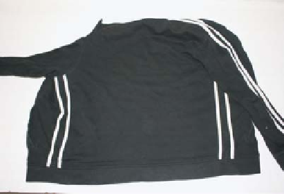
(a)
(b)
(c)
Figure 3.7
Location of saliva using Phadebas
paper. (a) Even with appropriate lighting the
identification of saliva stains can be difficult. (b) The article being examined is moistened using
sterile DNA-free water and Phadebas
paper is placed on top with the carbohydrate dye-coated
side in contact with the fabric; a glass plate holds the paper in contact. (c)
α
-amylase breaks down
the carbohydrate-dye complex, and the dye migrates through the paper and can be visualized