Biology Reference
In-Depth Information
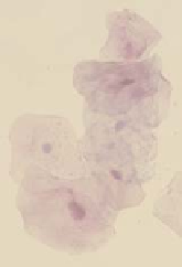
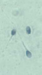
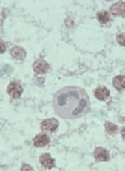
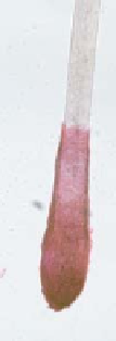
(a) (b) (c) (d)
Figure 3.2
Common cell types that are recovered from scenes of crime: (a) white blood cells;
(b) spermatozoa; (c) epithelial cells (from saliva); and (d) a hair shaft with the follicle attached.
(The cells have been stained with haematoxylin and eosin)
Collection and handling of material at the crime scene
The high level of sensitivity that makes DNA profiling an invaluable forensic tool can
also be a potential disadvantage. Contamination of evidential material with biological
material from another source, such as an attending police officer or scene of crime
officer, is a very real possibility. It is vital that the appropriate care is taken, such
as maintaining the integrity of the scene and wearing full protective suits and face
masks during the investigation of the scene [7 - 9] (Figure 3.3). Improper handling of
the evidence can have serious consequences. In the worst cases, it can cause cross-
contamination, lead to sample degradation and prevent or confuse the interpretation
of evidence.
Identification and characterization of biological evidence
Locating biological material is necessary before collection for further analysis can
occur. Furthermore, identification of the source of the material, for example demon-
strating that a stain is blood, can be a vital piece of information in a given case,
even before any DNA analysis is undertaken.
Searching for biological material, both at the crime scene and in the forensic labo-
ratory is performed primarily by eye. In the laboratory low-power search microscopes
may help to localize stains and contact marks. The use of either chemical or physical
methods can be used to detect biological materials. Alternative light sources (ALSs)
using both infrared and ultraviolet light can provide a contrast between the fluores-
cence of proteins in the body fluid and the background substrate. Chemical methods
use either the production of light or a colour change reaction. These techniques