Information Technology Reference
In-Depth Information
intuition, the diagnostic decision taken may be far from the best. The steps of the
visual assessment of the analysed image are frequently preceded by some manual
modifications of the image structure and its presentation method to improve the
ability to visually assess the specific case. A large number of computer tools have
been developed for this purpose and their operation is satisfactory [9][10][17].
However, there are no similar IT tools to be used for the automatic support of in-
tellectual processes of physicians when they analyse an image and interpret it.
Obviously this is not about eliminating humans from the decision-making process,
as this diagnostic stage requires the in-depth consideration by a physician, during
which he/she analyses the premises for the decision to be made, accounts for many
facts that the computer could not grasp, and he/she is also legally and morally re-
sponsible for the diagnosis made and the treatment undertaken based on it. How-
ever, at the stage of collecting premises for taking the decision, doctors should
make better use of the opportunities, offered by IT, to intelligently analyse com-
plex image data and attempt to automatically classify, interpret and even under-
stand specific images and their components using computer technology. So
far, there are no such intelligent IT systems to support the cognitive processes
of doctors who analyse complicated cases. However, it should be noted that
bolder and more productive attempts at developing them are being made
[1][10][12][20][23][24], including by the author. The reason for this is that there
are still a number of unsolved scientific and technical problems encountered by
designers of intelligent cognitive systems. What is more, these problems multiply
greatly if we move from computer programs supporting the analysis of data pro-
vided by relatively simple measuring devices to second generation IT systems
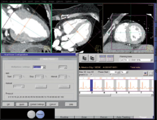
[6][8][15] coupled to medical diagnostic apparatuses, e.g. the very 3D images of
coronary vascularisation considered here (fig. 2.).
Fig. 2
The operator's panel of the computer system coupled to SOMATOM Sensation Car-
diac 64 (source: [4])
Search WWH ::

Custom Search