Information Technology Reference
In-Depth Information
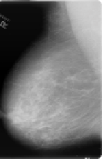
MLO-Right
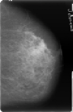
CC-Right
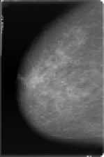
MLO-Left
CC-Left
Fig. 1.
MLO and CC projections of a left and right breast. The circle represents a lesion
and the irregular areas represent regions, true positive and false positive, detected by
a single-view CAD system
the most effective strategy on reducing mortality rates. This is the reason why
many countries have introduced breast cancer screening programs which target
at women of a certain age, usually starting at age of 50 years, aiming to detect
the occurrence of breast cancer as early as possible. Breast cancer screening is
widely seen as one of the keystone of breast cancer management, and an im-
pressive decrease in breast cancer mortality in the age group invited for breast
cancer screening has been shown [1].
The screening process involves carrying out an X-ray examination of the
breasts and the reading of the resulting images, or
mammograms
,bytrained
radiologists. A screening mammographic exam usually consists of four images,
corresponding to each breast scanned in two views - mediolateral (MLO) view
and craniocaudal (CC) view; see Figure 1. The MLO projection is taken un-
der (approximately) 45
◦
angle and shows part of the pectoral muscles. The CC
projection is a top-down view of the breast.
Despite the high level of expertise of radiologists involved in breast cancer
screening, the number of misinterpretations in the screening process is still high.
For instance, it is not unusual to encounter cases of women which have breast
cancer, but have their lesions missed during the reading process. This is due to
the fact that diagnosis is subject to one's interpretation: either a lesion repre-
senting the cancer is not detected by the radiologist or the lesion is identified
but incorrectly evaluated. In other words, mammogram reading is not a straight-
forward task: image quality, breast density and a vague definition of the char-
acteristics of breast cancer lesions are some of the factors which influence such
reading.
In order to support radiologists on their observations, and interpretation,
computer-aided detection (CAD) systems are used. Most CAD systems have
the purpose of helping avoiding detection errors. However, CAD systems can
also be exploited to increase the radiologist's performance by providing some
interpretation to mammograms [2].


Search WWH ::

Custom Search