Information Technology Reference
In-Depth Information
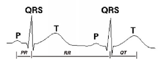
Fig. 1.
The main waves of an ECG.
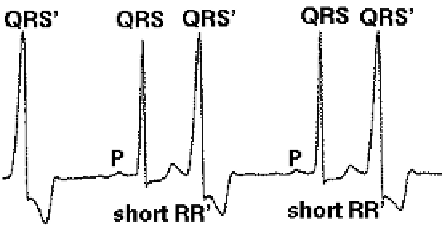
Fig. 2.
An example of arrhythmia: bigeminy.
to the bottom of the heart through the atrioventricular (AV) node and the right and left
bundle branches and back from the bottom to the top of the right and left ventricles.
The cardiac muscular cells located along the propagation path contract themselves at
the passage of the depolarization front and this causes the contraction of the atria and
the ventricles which in turn provokes the expulsion of the blood in the body. The elec-
trical activity displayed by ECG is generally described by a succession of characteristic
waves. In cardiology, these waves are labeled by letters starting from P. The most in-
formative are the
Pwave
which can be related to the atria activation, the succession of
the Q, R and S waves called the
QRS complex
which can be related to the ventricles
activation and the
Twave
which corresponds to the repolarization of the cardiac elec-
trical cells (cf. Fig. 1). Wave shapes and the time elapsed between successive waves are
major features from which the heart condition and different cardiac disorders can be in-
ferred. The succession P - QRS - T represents a normal cardiac beat and the succession
of normal cardiac beats constitutes a normal ECG.
A
cardiac arrhythmia
occurs when the electrical activity becomes abnormal due, for
example, to electrical problems on the propagation path. As a consequence the heart
rhythm may become faster, lower or irregular and wave shapes may change or some
waves are not present or extra waves appear. As an example, Fig. 2 displays an ECG
related to the bigeminy arrhythmia. In this disorder, an extra activation node, called
an ectopic node, located in one of the ventricles triggers extra heart contractions also
called premature ventricular contractions (PVC). The primed QRS complexes, noted
QRS', are related to this kind of extra activations.

Search WWH ::

Custom Search