Image Processing Reference
In-Depth Information
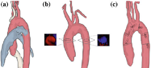
Fig. 25.5
Anatomical context visualization with cel shaded silhouettes and occluding contours.
a
Thoracic arteries.
b
MPR visualization exploded view of planes at cross-section positions.
c
Flow-
rate arrow-trails arrows at 280ms after the start of the cardiac cycle. [
46
] © IEEE Reprinted, with
permission, from IEEE transactions on visualization and computer graphics 16(6)
25.4.2 Localization of Anatomical Landmarks
The exploration of vascular structures and the embedded flow benefits strongly from
geometric descriptors that enable a carefully guided or constrained interaction. Such
geometric descriptors may also be used to decompose the relevant anatomy in mean-
ingful subregions to ease the exploration of complex flow data.
A widely used geometric descriptor is the vessel centerline, determined by a
skeletonization algorithm (see, e.g., [
19
]). The vessel centerline is often used in order
to move a cross-sectional plane that is always aligned perpendicular to the centerline,
presenting the maximum-sized area. In conventional vessel analysis packages, the
cross-sectional view displays the intensity values from the original image data, e.g.,
the CT Hounsfield values. In case of blood flow data, this strategy may be used to
present any scalar value derived from the flow or the flow data itself, e.g., by using
some glyph mapping. Van Pelt et al. [
46
] presented this cross-sectional visualization
approach for the main arteries (see Fig.
25.5
).
Better support for exploration tasks may be achieved by detecting and analyz-
ing further anatomical landmarks of a particular region. Once these landmarks are
identified, they may be used for labeling and for guiding movements in the complex
3D anatomy. The choice of such landmarks is specific for a particular anatomical
region. We describe and illustrate this principle for the specific example of cerebral
aneurysms. First, it is essential to understand
which
landmarks are actually important
to characterize the local vessel anatomy. Neugebauer et al. [
29
] questioned a couple
of neuroradiologists to draw cerebral aneurysms and extracted characteristic points
commonly used by them. The following points were deemed essential (see Fig.
25.6
).
the
dome point
of an aneurysm,
the (curved)
ostium plane
, where the blood enters the aneurysm, and
a so-called
central aneurysm axis
(the closest connection between the parent ves-
sel's centerline and the dome point)
Search WWH ::

Custom Search