Image Processing Reference
In-Depth Information
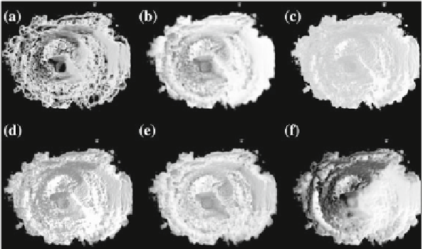
Fig. 24.8
Comparison of six volume shading models as applied to a 3D US scan of a human heart.
a
Phong,
b
[
62
],
c
[
76
],
d
[
37
],
e
[
66
],
f
[
44
]
account for scattering effects in slice-based volume rendering. Figure
24.8
eshows
the application of the directional occlusion shading technique [
66
]. This technique
constrains the light source position to coincide with the view point. Finally, Fig.
24.8
f
shows the application of a technique based on spherical harmonic lighting [
44
].
Advanced illumination techniques are now being implemented in the commercial
ultrasound workstations. Some workstations use additional color coding based on
depth. Deeper tissues are colored with cold tones such as blue while close regions
have red and orange tones. This effect has been firstly described by Einthoven [
14
]
and is also referred to as chromostereopsis [
2
]. Figure
24.7
a shows a chromatic depth-
encoding rendering of a 3D human heart in a modern ultrasound workstation.
24.7 Ultrasound and Augmented Reality
Ultrasound is commonly viewed on a separate monitor. Therefore, it is difficult to
comprehend the spatial relationship between what you see on the monitor and where
it is located in the patient's body. Augmented reality can aid the user by, for instance,
super-imposing the ultrasound image onto the body where the ultrasound probe is
positioned. Bajura et al. presented a system which linked 3D freehand ultrasound
with a head-mounted display (HMD) [
4
]. The HMD contains a camera, tracker and
two displays, one for each eye. The system can then project the tracked ultrasound
image onto the tracked camera feed so the user can see where in the body the image
is actually positioned.
Combining segmentation, surface rendering and augmented reality, Sato et al.
aimed to aid surgeons during breast
tumor removal for minimizing risk and
Search WWH ::

Custom Search