Biology Reference
In-Depth Information
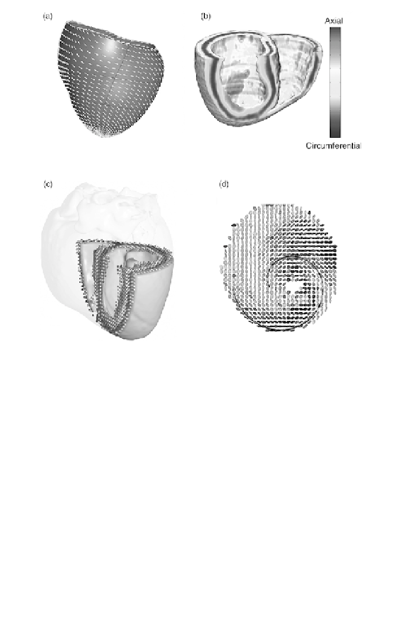
Figure 9.7
(a) Finite-element model of the canine ventricles with fiber
orientation displayed on the epicardial surface using short line segments.
(b) DTMRI-based reconstruction of fiber inclination angle (see color code)
throughout the canine ventricles. (c) Transmural variation of fiber inclination
angle (color coded as in (b)) with diffusion tensor data displayed using glyphs
(see text). (d) Spatial rotation of epicardial fibers near the apex of the heart.
Eigenvectors of the diffusion tensor define glyph orientation and
eigenvalues define glyph shape. In particular, cylindrical glyphs
indicate a single preferred direction of diffusion with diffusion being
isotropic in the transverse directions, and cuboid glyphs represent
orthotropic diffusion (three distinct eigenvalues indicative of myocar-
dial sheets or laminae). Figure 9.7d shows the spiral pattern that
cardiac fibers take near the apex of the heart as they plunge into the
infundibulum. Reconstruction of the high rate of twist of cardiac fibers
in this region is a challenging test of the spatial resolution of modern
DTMRI methods. These data demonstrate that DTMRI may be used
to reconstruct ventricular anatomy at high spatial resolution.
Search WWH ::

Custom Search