Biology Reference
In-Depth Information
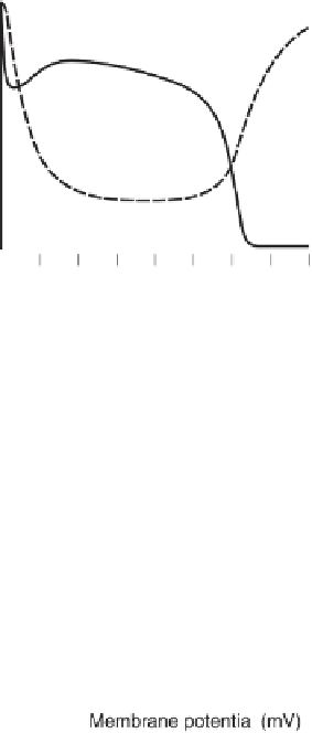
Figure 9.6
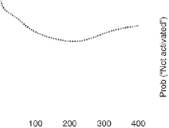
(a) Action potential (solid line) obtained in the CaRU model
when the fraction of LCCs not voltage-inactivated (dotted line) and not
Ca
2+
-inactivated (dashed line) is adjusted to match the experimental data of
Linz and Meyer [38]. Not stable AP, in contrast to those predicted when using
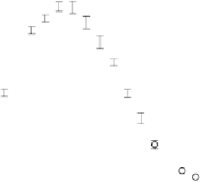
the common pool model in figure 9.5d. (b) Mean peak Ca
2+
flux amplitudes,
F
LCC(max)
(closed circles) and
F
RyR(max)
(open circles), as a function of membrane
voltage for the CaRU model,
n
=
5 simulations at each voltage.
Common pool models do incorporate biophysical detail but cannot
describe either graded CICR or predict stable APs. The CaRU model
described above is able to capture key experimentally measured
properties of CICR, but only at very high computational cost. A key
challenge that remains is to find simpler approaches to the integrative
modeling of CICR in cardiac myocytes that retain predictive power
while achieving reduced computational cost.


Search WWH ::

Custom Search