Agriculture Reference
In-Depth Information
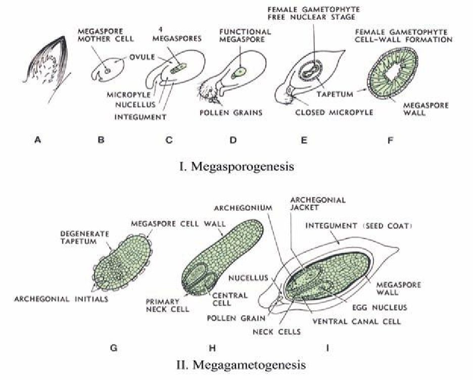
Figure 2.7. Ovule and female gametophyte development in Douglas ir (after Allen and Owens, 1972).
Pollination and fertilization
Pollen grains are formed within the microsporangia (pollen sacs) contained in the microsporophyll (male
spore-bearing organs) on the pollen cone. Pollen mother cells (microsporocytes) (2n) undergo meiosis
(chromosomal reduction division) forming four haploid microspores that following additional mitotic divi-
sions produce pollen grains which are shed as pollen from the male cone and are transported to the female
cone by wind.
The process of fertilization (Fig. 2.8) in gymnosperms involves only the union of the male gamete
from the pollen grain with the egg of the archegonium. This is in contrast to angiosperms which undergo
double fertilization resulting in a triploid endosperm and diploid zygote. In gymnosperms, the haploid
megametophyte functions as the nutritive tissue of the gymnosperm seed, similar to the function of endo-
sperm and perisperm in angiosperm seeds.
Another major difference exists between angiosperms and gymnosperms. In angiosperms, the time
interval between pollination and fertilization is generally a matter of hours or days. In gymnosperms, this
time interval may be several weeks or months. Douglas ir pollen does not fertilize the egg until ten weeks
after shedding, and is trapped within integuments of the ovule during this period. Many conifers, including
pines, have a 14-month interval between pollination and fertilization.