Agriculture Reference
In-Depth Information
SEroLoGy
Serological diagnostic techniques have long been used in human and veterinary pathology. They also can be
applied to detect pathogens in plant tissue. The technique utilizes antibodies generated during the immune
reaction in animals in response to invasion by alien materials (antigens). Antigens are proteins or carbo-
hydrate molecules that are components of microorganisms such as fungi, bacteria, and viruses. Antibodies
made in the animal against particular antigens are very speciic to the antigen. The antibodies are proteins
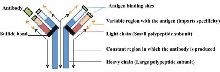
and have a basic structure as shown (Fig. 10.12). In an animal's immune system, antibodies bind with anti-
gens and expel them from the animal. Antibodies can be removed from animals either in blood serum or
by harvesting cells from the spleen. An antibody is capable of binding with the antigen, which it was made
against after it is extracted from the animal. This reaction can be used for diagnostic purposes.
Figure 10.12. Schematic diagram of the molecular structure of an antibody.
Antibodies to a plant pathogen are made by injecting an animal with a pure culture of the pathogen.
After a period of time, the animal will make a speciic antibody against the antigenic component unique
to the target pathogen. Before application in a diagnostic tool, the speciicity of an antibody to the target
pathogen is rigorously tested to ensure that it does not cross-react with other microorganisms (Lamka et al.,
1991).
A successful diagnostic system requires speciicity of the antibody and the ability to detect and quan-
tify the antibody-antigen complex. The latter can be achieved in several ways.
Agglutination.
Agglutination is the simplest technique available to detect antibody- antigen reactions.
The antibody and tissue extracts are mixed in solution. If the antigen is present, a precipitate will form in
the solution. If the antigen is not present, that solution will remain clear (Fig. 10.13).
Agar precipitin.
The most common form of agar precipitin tests is Ouchterlony double diffusion.
Filter paper disks, dampened with a solution of antiserum are surrounded by disks impregnated with ground
up seed tissue on agar gels (Fig. 10.14). The antibody and antigen in seed tissue diffuses from the respective
disks. A precipitin line in the agar between an antiserum disk and seed tissue disk indicates a reaction of the
antigen and antibody. No precipitin line between these disks indicates that the antigen was not present.