Biomedical Engineering Reference
In-Depth Information
a
3000
b
2500
15000
2000
10000
1500
1000
5000
Y
Y
500
X
X
0
0
25
c
25
d
20
20
15
15
10
10
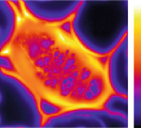
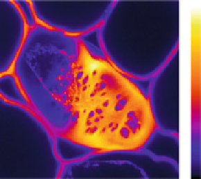
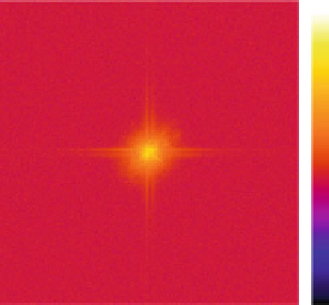
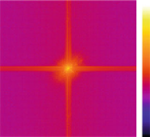
Fig. 4.3
Comparison between two fluorescence microscopes: Cross section through a
Convallaria
rhizome showing a blow up of a cortical parenchyma cell and its highly fluorescent wall (Courtesy
INRA). This sample was imaged on a (
a
) WFM (maximum intensity is 20,000 IU) and (
b
)CLSM
(maximum intensity is 3,000 IU) The intensities are linearly proportional to the number of photons
collected. (
c
) 2-D Fourier transform of the WFM image. (
d
) 2-D Fourier transform of the CLSM
image
have any protection against flux levels of 100-10
,
000 times higher than the normal
levels [
69
].
Although a CLSM is in principle a standard upright or inverted fluorescence
microscope equipped with a high quality objective lens, there are several fundamen-
tal differences between it and a WFM. CLSM has a smaller Depth-of-field (DOF)
[
70
], higher contrast, reduction of out-of-focus light, 'background rejection', and
full three-dimensional image scanning ability. In a conventional WFM, the entire
image is recorded onto the CCD camera. In contrast, in a CLSM, the specimen
is irradiated sequentially point-by-point using a laser beam as excitation source
and a pinhole that is “confocal” with this source. To be detected, the emitted
light must pass through this confocal aperture before being detected by a PMT.
Because fluorescence from out-of-focus planes will be out-of-focus at the plane of
the pinhole, most of it will not pass to the PMT with the result that the light that
is recorded comes from the optical section defined by the focus plane. The entire
specimen is scanned in 2-D or 3-D, generating a 2-D or 3-D image. There are many


Search WWH ::

Custom Search