Agriculture Reference
In-Depth Information
cell walls are composed of a complex matrix of
pectin, lignin and hemicellulose in which is
embedded an armoury of cellulose fibrils. The
arrangement and orientation of the cellulose
microfibrils in relation to the cell's axis allow
three closely associated zones to be identified
(Roland
et al
., 1995) (Fig. 3.4). Starting from
the outside and moving towards the lumen, the
zones are:
•
Secondary wall: this is put down in the
final stages of cell growth and is made
up of three layers, referred to as S1, S2
and S3.
The cellular types noted for their fibrous con-
tent show a predominance of rigid secondary
walls. The width, composition and architecture
of the different parietal layers will evolve
throughout the maturation of the plant cells
and vary according to the cell type, tissue type
and organ.
The middle lamella is rich in salts of pectic
acid, demonstrates hydrophilic properties in
young growing cells and contains little cellu-
lose. This layer glues adjacent cells together,
while the deposition of lignin further reinforces
this, providing stiffness and rigidity.
The primary wall is composed of cellulose
microfibrils embedded in a matrix of pectin and
hemicellulose. The cellulose fibres are widely
dispersed and aligned at all angles. This pari-
etal layer is generally lignified in fibrous plants.
The secondary wall is the main source of
fibres and is characterized by its high cellu-
lose content and the orientation of cellulose
microfibres in each of its three layers (S1, S2,
S3). In contrast to the primary wall, the
microfibres organize themselves in line with
the axis of the cell, while showing some vari-
ation between layers. In particular, the depo-
sition of cellulose microfibres is virtually
completely parallel to the cell axis in the thick
•
Middle lamella: forms the interface
between adjacent cells.
Primary wall: supple and extendable, this
•
zone allows cells to lengthen when young.
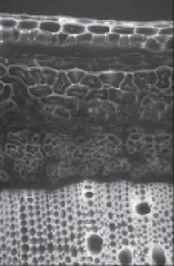
Epidermis
Primary
fibres
Secondary
fibres
Cambial zone
F
Xylem
5
50
50
μ
μ
m
m
V
Fig. 3.3.
Transverse section of a hemp stem.
Lignins are coloured using phloroglucinol and
hydrochloric acid. V = vessels; F = xylem fibres.
S3
Secondary
cell wall
S2
S1
Primary
cell wall
Middle
lamella
Fig. 3.4.
Architecture of the S1, S2 and S3 layers of the tracheid cell wall showing the relative orientation
of the cellulose microfibrils in each (from Brett and Waldron, 1996).