Biomedical Engineering Reference
In-Depth Information
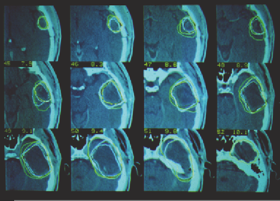
Figure 3.19. Delineation of the CTV on several sections of one
patient with two delineations at separate times by two radiation
oncologists (4 contours per section). Unpublished study by D.
Pontvert and N. Liebsch, MGH, USA.
In this exercise, two physicians drew a target volume on each of two
occasions on each of eight patients (only one of whom is represented
in Figure 3.19).
Automatic feature extraction
There is much research into what is termed “automatic feature
extraction.” Currently, good success is achieved for high-contrast
objects such as the external skin surface and outlines of the lungs,
both of which involve high-contrast tissue/air interfaces, and for bone
which is demarcated by the high contrast bone/soft tissue interface.
However, the majority of features have much lesser contrast relative
to their surroundings and, to date, there has been limited success in
the reliable automatic extraction of most features of interest.
I mentioned in the introduction to this chapter that the delineation of
the
tumor
is almost never possible by automatic means. The reasons
for this are stated in the introduction and there is no need to repeat
them here.