Biomedical Engineering Reference
In-Depth Information
shifted in range by a complex inhomogeneity. Rather, its slope can be
substantially less steep and less regular. There could be both a
significant underdose in a tumor, and a significant overdose in a
distal-lying normal tissue if this degradation was not appreciated, or
was ignored.
The degradation of the falling edge of the Bragg peak
is as much as
±
2 cm at point C of Figure 11.5. In a companion
experiment, looking at the Bragg peak degradation of carbon ions
passing through the abdomen, the degradation was even somewhat
greater - which was attributed to the effects of organ motion during
the long exposure needed to take the data.
pristine Bragg Peak:
pristine Bragg Peak:
A
A
A
A
B
B
B
B
C
C
C
C
A
A
A
B
B
B
C
C
C
spread-out Bragg Peak:
spread-out Bragg Peak:
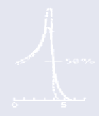
Figure 11.5. Degradation of a pristine Bragg peak (
right panel - top
) and
a spread-out Bragg peak (
right panel - bottom
) passing through a water-
filled human skull along paths behind three regions, A, B and C,
identified in the radiograph shown in the left panel (see text). The
unperturbed dose (i.e., when the skull is replaced by a water tank) is
shown as a dotted line in all panels. Reproduced with permission from
Urie
et al
. (1986a).
An uncertainty analysis (see Goitein (1985) and Chapter 8) can
establish the confidence limits on the dose distribution. Figure 11.6
shows an example of the computed bounds on the penetration of a
beam passing through the base of skull (Urie
et al
., 1986a). One sees
in this figure the calculated range uncertainty is greater in the shadow
of regions of complex heterogeneities, just as one would expect.
To cover the CTV to full dose at a given confidence level (the price
being that distal normal tissues receive a greater dose than desired)