Agriculture Reference
In-Depth Information
M
Va
L
*
Mv
M
NE
5
μ
m
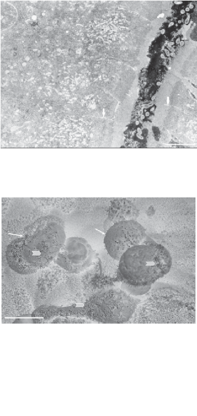
Fig. 3.2
Transmission electron microscopy micrograph of the anterior intestine (midgut) of rainbow trout
exposed to
V. anguillarum
(Va) in an
ex vivo
challenge. Clear signs of tissue damage are characterized by
necrotic enterocytes (NE) and disorganized microvilli (star). Key: L, lumen; M, mitochondria; Mv, microvilli;
arrow, tight junctional complexes. Scale bar
=
5
μ
m. (Source: Harper
et al
. 2011.)
F
10 n
Fig. 3.3
Scanning electron microscopy of the proximal intestine of Atlantic salmon exposed to
V.
anguillarum
. Note several detached or detaching enterocytes (arrows) lacking uniform microvilli at the
epithelial surface. Scale bar
=
10
μ
m. (Source: Ringø
et al
. 2007.)


Search WWH ::

Custom Search