Agriculture Reference
In-Depth Information
A
B
C
E
E
Lp
Lp
E
Bb
D
E
E
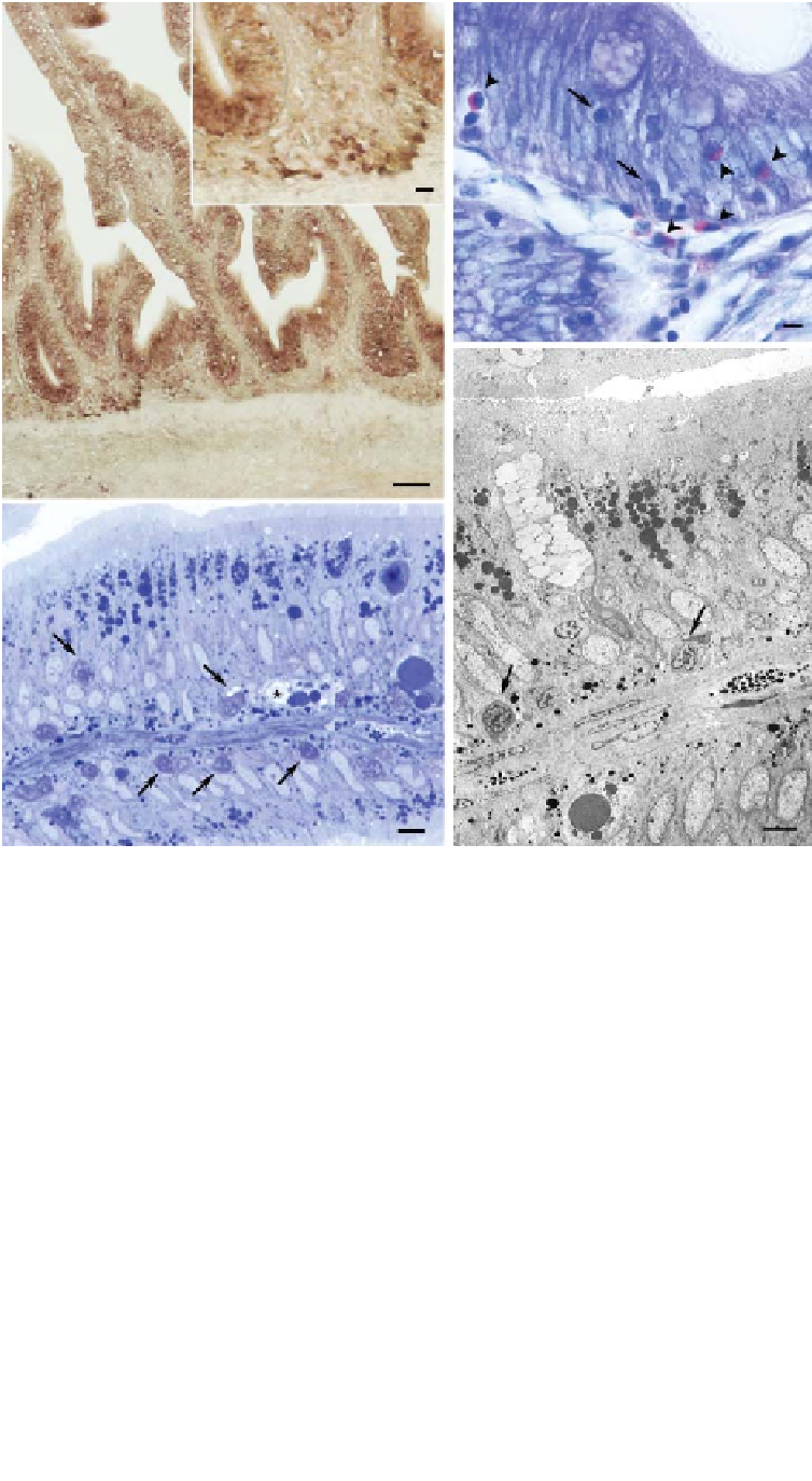
Fig. 2.1
Histological characterization of intestinal mucosal immune cells. Immune cells of the teleost
intestinal mucosa are less well organized and more diffusely arranged than in mammals. This figure shows
the distribution of some of these immune cells in the mucosa and the lamina propria of bony fish in a series
of five images. (A/B) Predominance of CD8
α
-positive cells in both the epithelium and the lamina propria of
the European sea bass intestine (CD8
α
RNA
in situ
hybridization). Scale bar
=
50
μ
m. The CD8
α
+
cells
aggregated in the lamina propria are shown at higher resolution in the insert (B), where the scale bar
=
10
μ
m. (C) May-Grümwald-Giemsa staining of the gilthead sea bream (
Sparus aurata
) intestinal mucosa,
showing lymphoid cells (arrows) and acidophilic granulocytes (arrowheads). Scale bar
=
4
μ
m. (D)
Semi-thin section of the gilthead sea bream intestinal mucosa housing numerous basolateral lymphocytes
(arrows) and an intraepithelial macrophage (star). Scale bar
=
5
μ
m. (E) Transmission electron microscopy
of the gilthead sea bream intestinal mucosa showing a goblet cell (star) amongst enterocytes and
basolateral lymphocytes (arrows). Scale bar
=
3
μ
m. Key: Ep, epithelium; Lp, lamina propria; Bb, brush
border. For colour detail see Plate 5.


Search WWH ::

Custom Search