Agriculture Reference
In-Depth Information
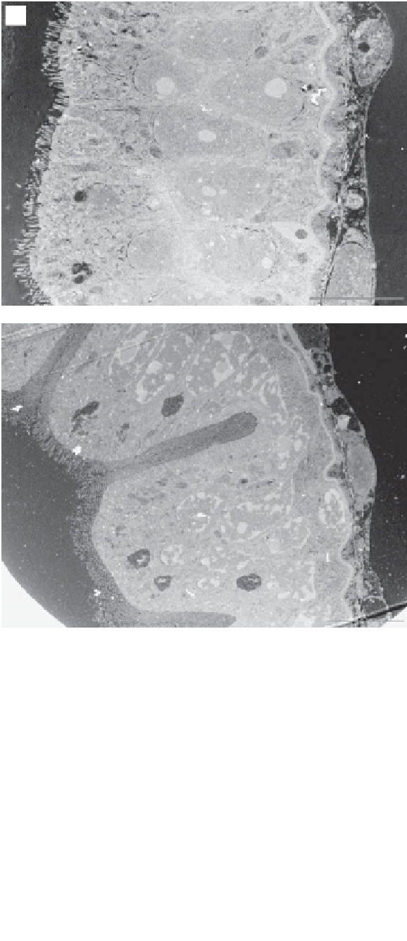
A
L
MV
MV
10 mm
B
N
L
N
10 mm
Fig. 15.1
Transmission electron micrographs of stage II larval lobsters showing the variation in the
degree of mucosal folding present at the apical brush border and the microvilli structure of (A) control fed
and (B) MOS fed individuals. Key: L, lumen; MV, microvilli; N, nuclei. Some stain precipitation is visible on
micrographs. Scale bar
=
10
μ
m. (Source: Daniels
et al.
2010.)


Search WWH ::

Custom Search