Agriculture Reference
In-Depth Information
A
B
G.G
G.C
C
D
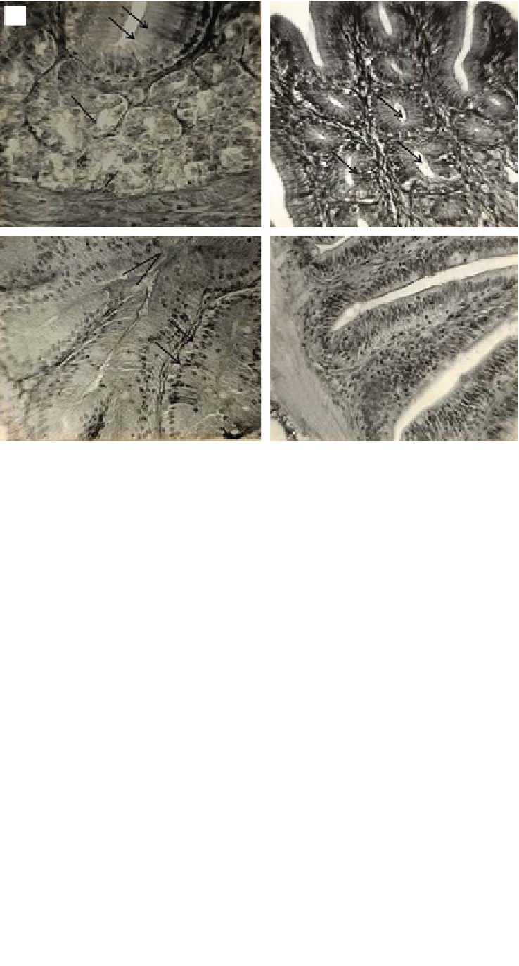
Fig. 1.3
Transverse section through different regions of the GI tract of climbing perch,
Anabas
testudineus
, a carnivorous perch. (A) Cardiac stomach. GG, gastric glands. Arrows indicate
mucus-secreting cells at the free borders of columnar epithelial cells.
×
400 magnification. (B) Pyloric
stomach. Arrows indicate tubular mucus glands or pyloric glands.
×
400 magnification. (C) The intestine.
GC, goblet cell. Arrows indicate absorptive cells.
×
400 magnification. (D) The pyloric caeca.
×
400
magnification. (Source: Ray and Moitra 1982.)
of gastric glands. The pH of the stomach therefore varies and in salmonids it is between 3.0
and 4.5 (Ransom
et al.
1984; Gislason
et al.
1996).
To our knowledge, the stomach microbiota is less investigated. Austin and Al-Zahrani
(1988) evaluated bacteria in the stomach of rainbow trout (
Oncorhynchusmykiss
Walbaum) by
using electron microscopy, while Navarrete
et al
. (2009) and Zhou
et al
. (2009a) evaluated the
stomach microbiota of Atlantic salmon (
Salmo salar
L.) and emperor red snapper (
Lutjanus
sebae
Cuvier), respectively, by molecular methods.
1.4 PYLORIC CAECA
In a number of fish species, several finger-like outgrowths develop from the anterior part of the
intestine in the region of pylorus. These are called pyloric caeca or intestinal caeca, and open
into the lumen of the intestine. They are located proximal in the midgut region, and, when


Search WWH ::

Custom Search