Biology Reference
In-Depth Information
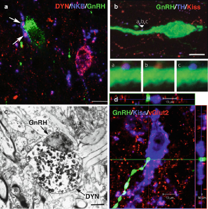
Fig. 3.2
Kisspeptin and KNDy connections with GnRH neurons. (
a
) KNDy inputs (
arrows
,
labeled with dynorphin and NKB) to a GnRH cell body in the mediobasal hypothalamus of the
sheep; a KNDy cell body (
magenta
) is seen nearby. Bar = 10 μm. Taken from Lehman MN, Coolen
LM, Goodman RL. Minireview: kisspeptin/neurokinin B/dynorphin (KNDy) cells of the arcuate
nucleus: a central node in the control of gonadotropin-releasing hormone secretion. Endocrinology
2010; 151(8):3479-3489 (with permission from The Endocrine Society). (
b
) RP3V kisspeptin
input (
arrowhead
, labeled with kisspeptin and TH) to a preoptic GnRH neuron in the mouse; (
a
) is
a higher power view of the contact, (
b
,
c
) show the same image with the TH (
b
) or kisspeptin
(
c
) channel removed. Bar=5 μm. Modifi ed from Clarkson J, Herbison AE. Dual phenotype
kisspeptin-dopamine neurones of the rostral periventricular area of the third ventricle project to
gonadotropin-releasing hormone neurones. Journal of Neuroendocrinology 2011; 23(4):293-301
(with permission from John Wiley & Sons). (
c
) Electron micrograph showing a dynorphin terminal
containing dense-core vesicles (
arrowheads
) in direct contact with a GnRH terminal in the sheep
median eminence. Bar = 2 μm. Taken from Lehman MN, Coolen LM, Goodman RL. Minireview:
kisspeptin/neurokinin B/dynorphin (KNDy) cells of the arcuate nucleus: a central node in the
control of gonadotropin-releasing hormone secretion. Endocrinology 2010; 151(8):3479-3489
(with permission from The Endocrine Society). (
d
) GnRH fi ber contacting a kisspeptin (KNDy)
cell in the ARC of the sheep; confocal orthogonal views through the close contact are shown above
and to the right. Section was also labeled for vGlut-2 showing co-localization in kisspeptin fi bers
and terminals. Bar = 10 μm. Unpublished data from Lehman, Cernea and Goodman, 2012
Search WWH ::

Custom Search