Biology Reference
In-Depth Information
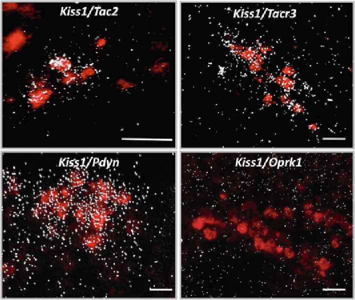
Fig. 15.3
Representative photomicrographs of mouse ARC illustrating the co-expression of
Kiss1
(labeled in
red
with digoxigenin coupled to
vector red
) and NKB (
Tac2
), NK3R (
Tacr3
), dynor-
phin A (
Pdyn
), and KOR (
Oprk1
) represented by
silver grains
. Scale bars = 50
μ
m
and the preoptic area (in mice and sheep, respectively) is virtually devoid of
these kisspeptin co-transmitters [
42
,
43
,
56
]. Consequently, it is reasonable to
infer that the role of NKB in the central control of reproductive function must be
related to the role of kisspeptin in the ARC, for example, in the negative feed-
back of sex steroids upon the gonadotropic axis.
An additional aspect in the physiology of NKB neurons that merits special atten-
tion is the identifi cation of the neuronal linage that generates the ARC population.
Recent work in male mice indicates that while the vast majority of
Kiss1
neurons in
the ARC seem to form a homogeneous population and collectively express
Tac2
[
56
], only approximately half of
Tac2
neurons in the ARC (at least in the male
mouse) appear to co-express
Kiss1
[
59
]. This fact demonstrates a subdivision of this
neuronal group with possible functional differences that yet remain to be unfolded.
To note, this phenomenon is in keeping with a previous description of two popula-
tions of NKB fi bers (with and without kisspeptin co-expression) in the median emi-
nence of female rats [
78
].
Search WWH ::

Custom Search