Biology Reference
In-Depth Information
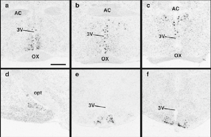
Fig. 13.1
The original photomicrographs detailing
Kiss1
mRNA distribution in the hypothalamus.
Silver grain clusters indicate areas where the labeled riboprobe is concentrated revealing
Kiss1
mRNA-expressing neurons. Cells were observed in the AVPV (
a
), periventricular nucleus (
b
),
anterodorsal preoptic nucleus (
c
), the medial amygdala (
d
), and the ARC (
e
,
f
).
3V
third ventricle;
AC
anterior commissure;
opt
optic tract;
OX
optic chiasm. Scale bar = 500
m. Data taken from
Smith JT, Dungan HM, Stoll EA, Gottsch ML, Braun RE, Eacker SM, Clifton DK, Steiner RA
2005 Differential regulation of KiSS-1 mRNA expression by sex steroids in the brain of the male
mouse. Endocrinology 146:2976-2984 with permission from The Endocrine Society
μ
cells were also noted in the anterodorsal POA, the medial amygdala, and the bed
nucleus of the stria terminalis (Fig.
13.1
).
Kiss1
mRNA localization in the AVPV
and ARC was also apparent in rats [
6
,
20
,
21
] and hamsters [
22
,
23
]. Confi rmation
of kisspeptin production in the AVPV and ARC was later provided with immuno-
histochemistry studies in mice [
12
,
24
], rats [
25
], and hamsters [
22
]. In sheep,
Kiss1
mRNA-expressing cells were shown to be located in the dorsolateral region of the
POA (perhaps a homologous population to the rodent AVPV/PEN) and the ARC
[
26
,
27
], with a similar distribution of kisspeptin-immunoreactive cells [
14
,
28
].
Populations of kisspeptin cells (mRNA and protein) in the POA and ARC have also
been reported in both human and nonhuman primates [
8
,
29
,
30
].
Kisspeptin Neurons Finding Their Way
If kisspeptin cells were critical in the feedback control of GnRH secretion, it would
be expected that kisspeptin neuronal fi bers make putative contacts with GnRH neuronal
cell bodies. In mice, kisspeptin-immunoreactive fi bers are located within the ventral
Search WWH ::

Custom Search