Biology Reference
In-Depth Information
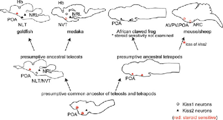
Fig. 2.4
Schematic illustrations for the distribution of
kiss1
and
kiss2
neurons in vertebrate brains,
including some hypotheses. Because
kiss1
and
kiss2
are duplicated paralogues, they are considered
to have been co-expressed in the same neurons in the common ancestor of teleosts and tetrapods.
Given that both amphibians and teleosts express
kiss2
in POA, the ancestral teleosts and ancestral
tetrapods should have expressed
kiss2
. Because
Kiss2
was lost in the mammalian lineage, we
hypothesize that
Kiss1
began to be expressed where
Kiss2
used to be expressed, to compensate for
the loss of
Kiss2
during mammalian evolution.
Open circles
indicate
kiss1
, and
fi lled triangles
indicate
kiss2
neurons.
Circles
/
triangles
in
red
are
kiss1
/
kiss2
neurons that are steroid sensitive
(fi gure as originally published in Kanda S and Oka Y (2012) Evolutionary insights into the steroid
sensitive
kiss1
and
kiss2
neurons in the vertebrate brain. Front. Endocrin. 3: doi:
10.3389/
hand, in goldfi sh, which lack
kiss1
neurons in NVT, POA
kiss2
neurons are the only
population of kisspeptin neurons that shows steroid sensitive kisspeptin mRNA
expression in the brain [
37
]. These POA
kiss2
neurons are also positively regulated
by gonadal steroids, as in the NVT
kiss1
neurons in medaka, and thus there has been
no report of negative regulation of kisspeptin expression in teleost brain so far.
Considering the report that positive or negative steroid feedback regulation can be
rather easily changed by the composition of co-expressing transcription factors
[
52
], the important common feature of the vertebrate kisspeptin neurons may be that
steroid sensitive kisspeptin neurons are localized in NVT and POA, which are
anatomically similar to arcuate and POA/AVPV in mammals, respectively. Although
the precise homology of brain nuclei between mammalian and nonmammalian
(especially teleost) kisspeptin neurons should be carefully discussed, the presence
or absence of sex steroid sensitivity in each nucleus may be one of the strongest
pieces of evidence to argue such homology.
An evolutionary working hypothesis of kisspeptin neuronal systems in vertebrates
is shown in Fig.
2.4
. In this hypothesis,
kiss1
- and
kiss2
-expressing neurons are dif-
ferentially distributed in the brains of mammal and other vertebrates. It is suggested
that the loss of
Kiss2
gene during mammalian evolution has been probably been
Search WWH ::

Custom Search