Biology Reference
In-Depth Information
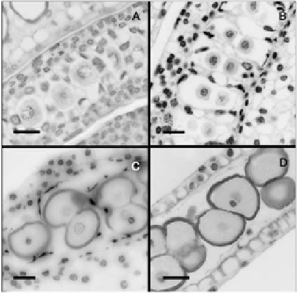
PMC (Figure 7A). After meiosis, the tapetum showed a vacuolization and a pro-
gressive degeneration as the tapetal chamber enlarged (Figure 7B). The nuclei of
the tapetal cells displayed elongated and lobular shapes together with a extremely
high chromatin condensation, revealed by an intense 4',6-diamidino-2-phenylin-
dole (DAPI) fluorescence (Figure 7C), typical features of programmed cell death
[28], which have also been found in the tapetal nuclei of other species [29]. At
the same time, tapetal cells released their cellular contents that coated the pollen
grains to form the pollenkit. At anther dehiscence the tapetum was completely
degenerated and had disappeared (Figure 7D).
Figure 7.
Tapetum degeneration in Annona cherimola.
(A) Pollen
mother cells in Prophase I and an active tapetum. (B) Dyad phase in enlarged tapetal chambers.
(C) Anther, 4 days before anthesis, showing bicellular pollen and degenerated tapetum, with nuclei displaying
elongated shapes and chromatin condensation. (D) Tapetum has disappeared in anthers of flowers at the female
stage showing mature pollen. Longitudinal 5
µ
m resin sections stained with DAPI. Bar = 20
µ
m.
Discussion
Pollen Development
Pollen in A. cherimola is shed in groups of four, originating from the same meiotic
division and, hence, the same tetrad. Pollen development, however, continues be-
yond tetrad formation and, although held together, pollen grains are fully mature
upon anther dehiscence. Meiosis cytokinesis occurred through the formation of
ingrowths of callose that are also found in genera of some primitive angiosperms
[30,31] including species of the Magnoliid clade as Magnolia tripetala [32] and