Biology Reference
In-Depth Information
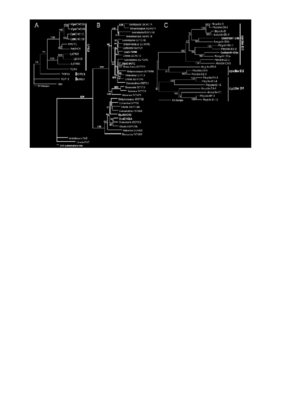
Figure 2.
Neighbor-joining trees of proteins encoded by CYC-like and D-type cyclin genes.
A) Neighbor-joining
tree of proteins encoded by the ECE lineage genes in CYC/TB1 subfamily, showing that
OpdCYC1C/1D and OpdCYC2A/2B form a branch that is sister to AmCYC/AmDICH from Antirrhinum
majus, which belong to the CYC2 clade in the ECE lineage. B) Phylogram of GCYC, showing the phylogenetic
relations of OpdCYC genes with other GCYC in Gesneriaceae. C) Neighbor-joining tree of proteins encoded
by D-type cyclin genes, showing that OpdcyclinD3a and OpdcyclinD3b are clustered with cyclinD3a and
cyclinD3b clades, respectively, in the cyclinD3 lineage. For sequence information see Methods. Phylogenetic
analyses were conducted using PAUP*4.0b4a, and bootsrap values over 50% (1,000 replicates) are indicated for
each branch.
Gene mRNA Expression Patterns
To assess the potential role of CYC-like genes in floral development, we conducted
in situ hybridization complemented by gene-specific RT-PCR on O. dinghushan-
ensis. As petal and stamen primordia began to emerge, OpdCYC1C mRNA was
detected in all five petal and stamen primordia (Figure 3A). Weak mRNA signals
were also detectable in the lateral edges and vascular tissue of sepals (Figure 3A).
After primordial initiation of petals and stamens, OpdCYC1C expression signals
were gradually weakened in the two lateral stamens (Figure 3B-D) with weak
mRNA detected in the ring meristem of the corolla-tube outside the stamen pri-
mordia (Figure 3B-C). Figures 3C and 3D were the successive sections from the
same individual flower across the base of stamen primordia (3C) and over their
upper parts (3D), respectively, which showed a size reduction from the base to the
upper part of the dorsal and ventral stamens. The mRNA signal of OpdCYC1C
was weak in lateral stamens while strong in both dorsal and ventral staminodes
in which its mRNA signal was stronger in the upper part than at the base (Figure
3C-D). OpdCYC1C mRNA also accumulated less in lateral petals than in dorsal
and ventral petals (Figure 3D). As the lateral stamens enlarged laterally, weak