Chemistry Reference
In-Depth Information
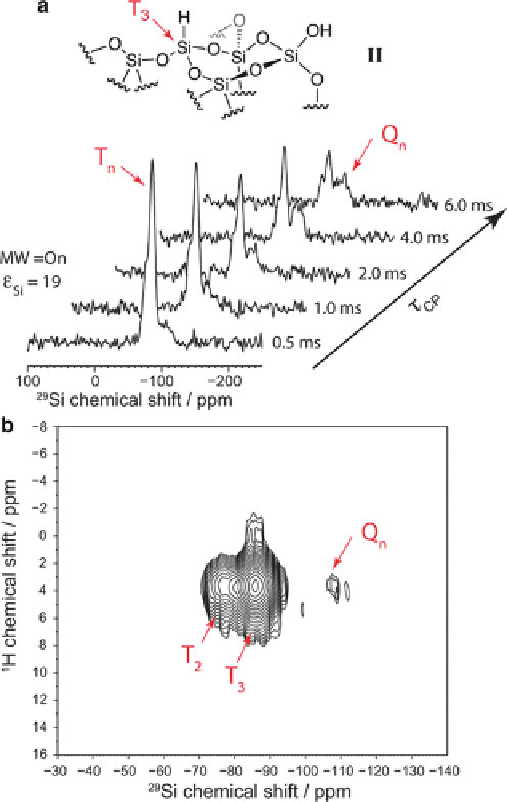
Fig. 6 (a) DNP-enhanced silicon-29 CPMAS spectra of compound II as a function of the CP
mixing time
CP
.(b) Contour plot of a two-dimensional
1
H-
29
Si spectrum of II recorded with DNP.
Reproduced with permission from [
49
]
t
that signal areas are not affected by different relaxation rates, and the method yields
chemical information on the detected molecules in the absence of sample derivati-
zation or sample separation. A principal limitation of conventional
1
H NMR is,
however, the relatively low signal dispersion over a spectral window of approxi-
mately 10 ppm, which restricts the capability to resolve chemical compounds in the
complex spectral backgrounds of biofluids. The use of
13
C NMR spectroscopy can
address poor signal dispersion problems as the
13
C chemical shift dispersion is
approximately 20-fold larger than that for
1
H. Spectral interference of the biofluid