Chemistry Reference
In-Depth Information
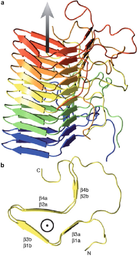
Fig. 5 Solid-state NMR structure of the HET-s (218-289) fibril. (A) Side view of the five central
molecules of the lowestenergy structure of the HET-s(218-289) heptamer calculated from the
NMR restraints. (B) Top view of the central molecule from (A). beta3 and beta4 lie on top of beta1
and beta2, respectively. Adapted from [
42
] with permission from the American Association for the
Advancement of Science
C13-C14-C15 regions (Fig.
6
). The large change at C13, C9, and C17 may be
attributed to the break of the Glu113 salt bridge, van der Waals contact, and local
hydrophobic environmental changes due to the displacements of the helices upon
activation. In addition to the studies on the retinal in rhodopsin, the structure of