Chemistry Reference
In-Depth Information
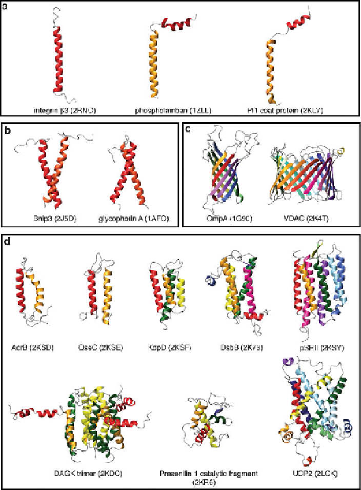
Fig. 6 Representative samples of integral membrane protein structures solved using solution
NMR,comprisedofeither(a) a single TM helix, (b) a TM helix dimer, (c)a
-barrel, or
(d) multiple TM helices from a single chain (with the exception of DAGK, which is a trimer of
3-TM helix subunits). Coloring from the N terminus to C goes from
red
to
orange, yellow,
green, blue, purple,
and then
pink
. All PDB accession numbers are indicated in parentheses for
each structure
b