Biology Reference
In-Depth Information
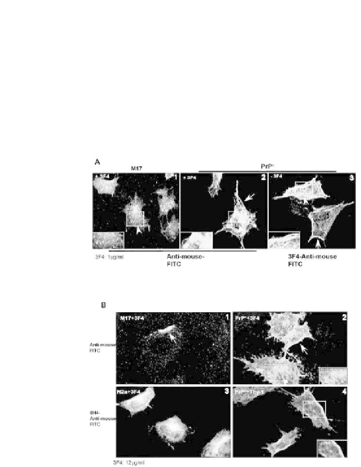
Exposure to 12 μg/ml of 3F4, however, cross-links PrP
C
at the plasma membrane and
reduces its endocytosis signifi cantly (Figure 6B, panels 1 and 2). As a control, mouse
neuroblastoma cells (N2a) expressing mouse PrP that does not react with 3F4 were
exposed to 3F4 and reacted with mouse PrP-specifi c antibody 8H4 followed by anti-
mouse-FITC. Examination shows normal distribution of PrP
C
at the plasma membrane
and some reactivity in the Golgi region as expected (Figure 6B, panel 3) [26]. Expo-
sure of PrP
C
cells to anti-Thy-1, a monoclonal antibody to an irrelevant GPI-linked
protein abundant on neuronal cells shows normal distribution of PrP
C
when reacted
with 8H4-anti-mouse-FITC (Figure 6B, panel 4), confi rming the specifi city of 3F4
mediated endocytosis and cross-linking of PrP
C
.
Figure 6.
Exposure of PrP
C
-cells to different concentrations of 3F4 induces endocytosis or cross-
linking of PrP. (A) Immunostaining of M17 and PrP
C
cells exposed to 1 μg/ml of 3F4 for 5 days
shows a prominent reaction in vesicular structures in M17 and PrP
C
cells (panels 1 and 2). Coalesced
vesicles simulating aggregated PrP
C
are evident near the Golgi region and in the cytosol of PrP
C
cells (panel 2). Untreated PrP
C
-cells reacted with 8H4-anti-mouse-FITC show a prominent reaction
at the plasma membrane as expected (panel 3). (B) Reaction of M17 and PrP
C
cells exposed to 12
μg/ml of 3F4 for 4 hr with anti-mouse FITC shows cross-linking of PrP on the plasma membrane of
M17 and PrP
C
cells (panels 1 and 2, arrow) and a slight increase of reactivity in vesicular structures
in the latter (panel 2, arrow-head). Similar exposure of N2a-cells to 3F4 and PrP
C
-cells to anti-Thy1
antibody followed by immunoreaction with 8H4-anti-mouse-FITC shows plasma membrane and
Golgi reaction of endogenous PrP in N2a cells (panel 3) and plasma membrane distribution of PrP in
Thy-1 exposed cells (panel 4). (Mouse PrP expressed by N2a cells does not react with 3F4).