Biology Reference
In-Depth Information
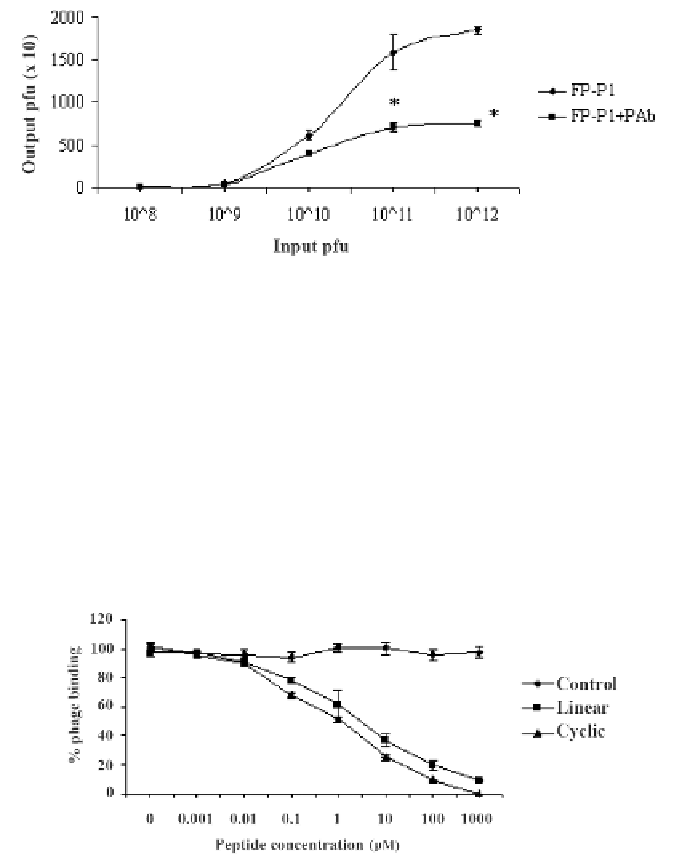
the number of phages bound to the AIV coated wells reduced dramatically as a result
of the competition between these two molecules for the same binding site on AIV.
For example, at input pfu of 1 × 10
12
/100 μl, the output pfu for the FP-P1 phage alone
was 1.8 × 10
4
plaques but in the presence of pAb, the output was reduced to 7.5 × 10
3
plaques, which is almost 2.4-fold reduction. This result clearly shows us that the phage
molecules that display peptides on their surface can compete for the epitope binding
sites on AIV with polyclonal antibodies.
Figure 7.
Antibody-phage competition assay. The phage competes with polyclonal antibodies for
binding site on AIV, suggesting they may share common binding sites. Experiments were done in
triplicates and the error bars represent the standard deviation of the mean. *, statistical significance
(P < 0.05).
Peptide-phage Competition Assay
In order to identify whether the synthetic peptides and the phages (FP-P1) compete
for the same binding sites on AIV H9N2, a peptide-phage competitive assay was per-
formed. When the peptides (both linear and cyclic) were pre-incubated with the virus,
the number of phages bound to the virus was reduced gradually in a dose-dependent
manner. At 1 mM concentration of the peptides, the phage binding was almost com-
pletely inhibited (Figure 8). The control peptide does not possess any inhibitory effects
on phage binding to AIV.
Figure 8.
Peptide-phage competition assay. The peptides competes with the fusion phage FP-P1 for
binding sites on AIV, suggesting that peptides displayed on the fusion phage FP-P1, and not other
parts of the phage, binds to the AIV. Experiments were performed in triplicates and the error bars
represent the standard deviation of the mean.