Biology Reference
In-Depth Information
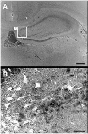
Figure 6.
Survival of grafted F3.Akt1 human NSCs in hippocampus was demonstrated by immunoperoxidase
microscopy of hNuMA at 8 weeks post-transplantation (PT).
A: Lower magnification of hippocampus 8 weeks
PT. Bar indicates 1 mm. B: F3.Akt1 human NSCs grafted in cortex overlying striatum were found to migrate
extensively to hippocampus 8 weeks PT. Bar indicates 50 µm.
To determine whether Akt1 expression in F3.Akt1 cells induces their own
proliferation, expression of cell proliferation marker Ki-67 was examined in ICH
brain sections. Transplanted F3.Akt1 cells identified as hNuMA-positive cells
were immunoreaction-negative for proliferation marker Ki-67 indicating that the
grafted F3.Akt1 cells did not continue to proliferate following transplantation.
Furthermore there was no sign of tissue distortion or tumor formation in brain
of ICH animals grafted with F3 or F3.Akt1 hNSCs 6 months PT. Good survival
of hNSCs was found in the hNSC injection path into striatum two days after
transplantation.
discussion
In the present study, mouse ICH model was used to provide proof-of-principle
that hNSCs genetically modified to express Akt1, a general mediator of cell sur-
vival signals, can be transplanted in the brain of animal models of neurological
diseases, and produces beneficial effects of increased survival of grafted NSCs and
consequent functional recovery. We demonstrate that brain transplantation of
hNSCs overexpressing Akt1 in the collagenase-induced ICH mice resulted in im-
provement in motor performance as determined by rotarod and limb placement

Search WWH ::

Custom Search