Biology Reference
In-Depth Information
transplanted nscs differentiate into neurons and Astrocytes
At 7 days after induction of experimental ICH, 2×10
5
cells/2 µl of F3 or F3.Akt1
hNSCs were transplanted into ICH mouse cerebral cortex overlying hemorrhage
lesion site, 2 mm cranial to the hemorrhagic lesion. LacZ+ human NSCs mi-
grated selectively to the hemorrhagic core and also located on the border of the
hemorrhagic core and further away from the injection sites (Figures 4A-C). A
large number of transplanted hNuMA (human specific nuclear matrix antigen)-
positive F3.Akt1 cells (35-45%) differentiated into NF-H+ neurons in the peri-
hematomal sites (Figures 4D-F). While only a small number of transplanted
hNuMA+ F3.Akt1 cells (~4%) were GFAP+ astrocytes and the hNuMA+/GFAP+
double-positive cells were found along the border of hemorrhagic core (Figures
4G-I). These results indicate that a large portion of grafted F3.Akt1 cells dif-
ferentiate into either neurons or astrocytes in response to signals from the local
microenvironment provided by the hemorrhagic lesion.
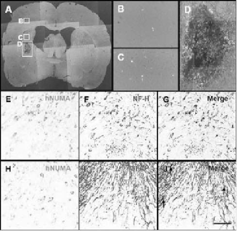
Figure 4.
LacZ (beta-galactosidase)-labeled F3.Akt1 human NSCs in intracerebral hemorrhage (ICH) mouse
brain at 2 weeks post-transplantation.
A: One week after an ICH lesion (an intrastriatal injection of collagenase),
LacZ -labeled F3.Akt1 NSCs were transplanted into the cortex overlying the ICH lesion. Two weeks post-
transplantation, LacZ-positive F3.Akt1 NSCs were found to migrate extensively into the hemorrhage core and
surrounding brain sites. Bar indicates 50 µm. B-D: Higher magnification of indicated areas. Bar indicates 20
µm. E-G: Double immunofluorescent staining of engrafted F3.Akt1 human NSCs in ICH mouse brain 8
weeks post-transplantation. F3.Akt1 NSCs in the lesion sites are found to differentiate into neurons as shown
by double staining of human nuclear matrix antigen (hNuMA) and neurofilament protein (NF-H, a neuron
specific marker). H-J: F3.Akt1 human NSCs in the lesion sites are found to differentiate into astrocytes (arrows)
as shown by double staining of human nuclear matrix antigen (hNuMA) and glial fibrillary acidic protein
(GFAP, an astrocyte specific marker). Bar indicates 20 µm.

Search WWH ::

Custom Search