Biology Reference
In-Depth Information
Figure 5.
Bromodeoxyuridine-labeling index (number labeled per 500 cells) in hormone-treated p53-
null mammary epithelial transplants. Transplants from each experiment (1, 2 and 3) were assayed for
bromodeoxyuridine. The black bars represent untreated transplants, the gray bars hormone-treated transplants.
The number above each pair of bars indicates the number of weeks after the removal of hormones. In experiment
3, 12 weeks after the removal of hormone represents 8 weeks after transplantation. There were four transplants
per treatment group. Five hundred cells were counted in each transplant. All four comparisons were significantly
different (p < 0.05).
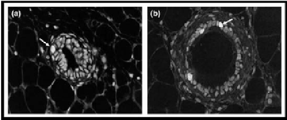
Figure 6.
Immunohistochemistry of bromodeoxyuridine-labeled mammary epithelial cells. The nuclei (arrows)
represent the uptake of bromodeoxyuridine into cells undergoing DNA synthesis. (a) Untreated; (b) treated with
estrogen plus progesterone.
Effect of Hormone Treatment on MMTV-Neu Mammary
tumorigenesis
The marked response of the p53-null mammary epithelium to a short-term expo-
sure to hormone raised the question of whether a similar response would occur
in a more tumorigenic model of mammary tumorigenesis. The MMTV-activated
neu model provided such a model, because tumors develop rapidly and with high
multiplicity. The effect of either short-term treatment with estradiol or estradiol
plus progesterone on mammary carcinogenesis was tested. The data in Figure 7
and Table 1 indicate that both treatments were equally effective in providing pro-
tection. Mammary cancer incidence after treatment with estradiol alone (33%) or


Search WWH ::

Custom Search