Information Technology Reference
In-Depth Information
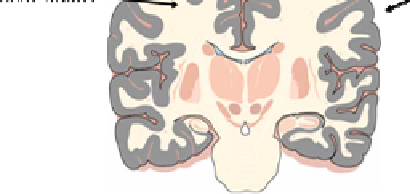
Fig. 2.
Gray matter was used to isolate the brain microvascular endothelial cells in the experiment
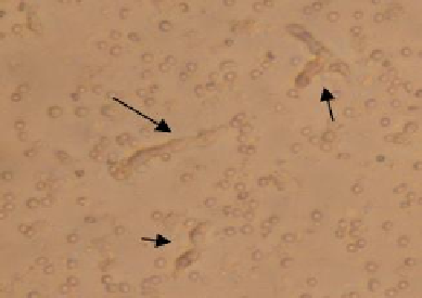
Fig. 3.
The isolated microvessels after digesting: digested using Collagenase/dispase and
DNase
Ⅰ
at 37
℃
for 30min, till most microvessels beaded and shorter then before.
(
200
×
)
The pellets was resuspended and dissociated with 2ml digestive juice, at 37
℃
for
30min, or the digestive suspension would be observed using microscope every 10min
from 20th minute on, till most microvessels beaded and shorter then before (Figure 3).
The microvessels were centrifuged at 150g, room temperature for 5min, and discarded
supernatant. Then the pellet was resuspended with PBS and centrifuged at 150g, room
temperature for 5min, twice. Then the pellet was resuspended with culture medium,
plated in collagen-coated 25cm
2
flask (Greiner, Bio-one, Germany), 1.5ml/flask,
incubated in a water-saturated atmosphere of air and 5%CO
2
. 24 hours later, when the
cells had adhered, the culture medium should be changed every other day, until the
cells became a confluent monolayer. Then the cells could be freezing or passage.
3 Results
3.1 BMEC Were Cultivated in 24 Hours
The collagen-coated surface is very beneficial for adherence of microvessels, micro-
scopically capillaries and a few red cells could be observed. After 5 to 6 hours of


Search WWH ::

Custom Search