Information Technology Reference
In-Depth Information
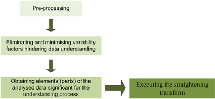
Fig. 2.21.
Pre-processing in the process of understanding the analysed medical image
The presented approach to the analysis is a concept aimed at the semantic inter-
pretation of medical images [87], [92], [97].
Pre-processing produces images cleaned of noise and reduced to such an extent,
in the sense of eliminating superfluous details, that they become graphs of
'straightened' structures, which are then approximated using an adaptively se-
lected broken line.
The above one-dimensional graphs mainly present information on the variabil-
ity of the width and the profile of the edge of the organ in question. These graphs
are used for understanding the contents of the analysed images under the assump-
tion that in the medical problem under consideration this form of representation is
sufficient to extract features of the image that are diagnostically significant. Obvi-
ously this may not be true for every type and form of images. However, this type
of an analysis has found prior application to the automatic interpretation of a
broad range of images from diagnostic examinations of organs in the abdominal
cavity, the chest and the vascular (circulatory) system [87], [112]-[117], so there is
every reason to also use it for pre-processing images of the spinal cord, bones of
lower and upper extremities.
It is important to adapt linguistic methods of the cognitive analysis of medical
images by generalising them for the structure of the nervous system and the skele-
ton of feet and hands, as the linguistic approach supports analysing morphological
changes in the shape of anatomical structures of the spinal cord and bones of ex-
tremities. In addition, using grammars of sufficient generative strength also makes
it possible to identify additional information (mainly semantic in nature) charac-
terising the organ or structure examined.